
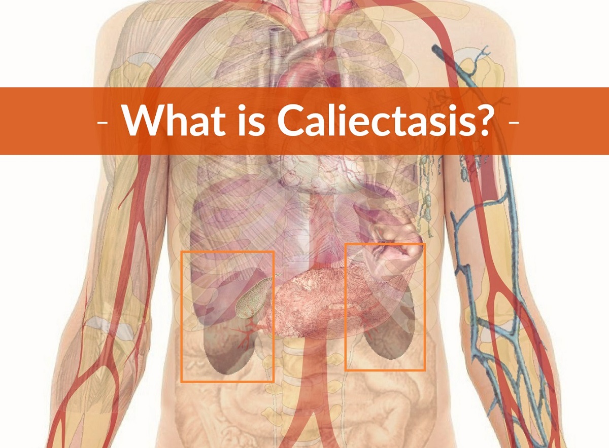
Caliectasis (hydrocalycosis) is a medical term used when the kidney’s calyces become stretched or dilated. In plain English, the small cup-like areas of the kidney that collect urine look larger than they should.
The calyces are part of the kidney’s drainage system, collecting urine before it moves into the renal pelvis, then down the ureter and into the bladder. Older medical references, including the Concise Medical Dictionary and Encyclopedia.com, define hydrocalycosis in this same general way.
What matters most, though, is that caliectasis is usually not the final diagnosis. It is often a clue on imaging that something may be slowing, blocking or changing the normal flow of urine.
That “something” could be temporary and mild, or it could be more serious. Annoying answer, I know, but kidneys like details.
A person with caliectasis may be evaluated by a primary care doctor first, but a nephrologist or urologist is often involved, especially if kidney function is affected or a blockage is suspected. The main job is to figure out why the calyces are dilated and whether urine needs to be drained more urgently.
Caliectasis Causes
There are several reasons the calyces can become dilated. Most commonly, doctors think first about obstruction somewhere along the urinary tract, because urine that cannot flow normally may back up toward the kidney.
As explained by the Merck Manual, urinary tract obstruction can occur in the kidney, ureter, bladder or urethra. MedlinePlus also notes that swelling of the kidney from backed-up urine is often related to blockage or reflux.
Possible causes include:
» Kidney stones blocking urine flow;
» A blood clot in the urinary tract;
» Narrowing or scarring of the ureter, sometimes after surgery, radiation or injury;
» Enlarged prostate pressing on the urinary outlet in men;
» Tumors or masses in the bladder, kidney, pelvis or abdomen;
» Urinary tract infection causing swelling or inflammation;
» Vesicoureteral reflux, where urine flows backward from the bladder toward the kidneys;
» Neurogenic bladder, where nerve problems affect bladder emptying;
» Severe constipation or fecal impaction pressing on the urinary tract;
» Congenital urinary tract differences present from birth.
Pregnancy can also cause dilation of the kidney collecting system, especially on the right side. In many pregnant patients this is a physiologic change, meaning it happens because of pressure and hormonal changes rather than a dangerous disease process.
That said, pregnancy does not make urinary symptoms something to ignore. Pain, fever, chills, vomiting or blood in the urine still needs medical attention.
Doctors also have to be careful not to overcall caliectasis. A radiology review in RadioGraphics notes that normal structures, such as prominent renal pyramids, may sometimes be mistaken for dilation, particularly in children and younger adults.
Symptoms
Caliectasis itself may not cause symptoms. Many people only learn about it after an ultrasound, CT scan or other imaging test is done for another reason.
When symptoms do happen, they are usually related to the underlying cause. For example, a kidney stone can cause severe flank pain, while a urinary tract infection may cause burning, urgency or fever.
Possible symptoms include:
» Pain in the back, side or flank;
» Tenderness over one or both kidneys;
» Difficulty urinating or feeling unable to empty the bladder;
» Urinating less than usual;
» Blood in the urine;
» Fever or chills, especially with infection;
» Nausea or vomiting with severe pain;
» Swelling or abdominal discomfort in some cases.
A urinary tract infection, kidney infection, stone or obstruction can look similar at first. This is why doctors usually do not rely on symptoms alone.
Caliectasis Diagnosis

The first test is often an ultrasound because it is noninvasive and does not use radiation. It can show whether the kidney collecting system looks dilated and whether one or both kidneys are involved.
In some cases, the doctor may order a CT scan, CT urography, MRI or an intravenous urography test. These tests can help show whether there is a stone, tumor, stricture or another reason urine may not be draining well.
Blood and urine tests are also important. A CBC may be used to look for signs of infection, while blood chemistry tests can check kidney function and electrolyte balance.
The doctor may order testing to look for:
» A high white blood cell count, which may suggest infection;
» Elevated creatinine or blood urea nitrogen, which may suggest reduced kidney function;
» Electrolyte abnormalities, especially if vomiting, dehydration or kidney dysfunction is present;
» Blood, protein, bacteria or crystals in the urine.
A urinalysis test can help detect blood or infection even when the urine looks normal. Sometimes people do not see visible blood, but the lab still finds red blood cells under the microscope.
For children and babies, the workup may be a little different. Pediatric specialists may use ultrasound follow-up, voiding cystourethrogram or nuclear medicine studies, depending on whether reflux, obstruction or congenital hydronephrosis is suspected.
Urinary Tract Obstruction or Caliectasis
Caliectasis and hydronephrosis are related, but they are not always used in exactly the same way. Caliectasis refers to dilation of the calyces, while hydronephrosis usually describes swelling of the kidney collecting system more broadly because urine is backing up.
In real-world doctor language, the terms can overlap. That is why the report matters less than the next question: what is causing it?
As described in “Differential Diagnosis in Abdominal Ultrasound” (Bisset et al., 2008), diagnosing caliectasis requires both confirmation and exclusion of other conditions. In other words, the doctor is not just looking for enlarged calyces; the doctor is looking for the reason.
When urinary tract obstruction is being considered, testing may help determine whether:
» The dilation is mild, moderate or severe;
» One kidney or both kidneys are affected;
» A stone is blocking the ureter;
» The obstruction is acute, intermittent or chronic;
» Bladder retention is causing urine to back up;
» A staghorn calculus or large stone is obscuring the collecting system;
» There is reflux instead of a true blockage.
Technical factors can make diagnosis harder, including obesity, bowel gas, dehydration or limited visibility during ultrasound. Not every unclear scan means something terrible is happening, but it does mean follow-up may be needed.
Pregnant women may have dilation of the renal collecting system, often most noticeable later in pregnancy. Older teaching from Harvard Medical School also describes this right-sided predominance, which doctors still commonly recognize in practice.
In unborn babies, hydronephrosis may be detected on routine prenatal ultrasound. Boston Children’s Hospital notes that hydronephrosis is one of the most common urinary tract findings seen before birth.
After birth, babies and children may be monitored with repeat imaging or referred to pediatric urology. Blood in the urine, kidney swelling, fever, poor feeding or pain can lead to further testing.
Treatment

Treatment depends on the cause, severity and whether kidney function is at risk. There is not one single “caliectasis pill,” which would be convenient, but unfortunately kidneys did not ask us.
The goal is usually to treat infection if present, relieve obstruction if present and protect kidney function. Options may include:
» Observation and follow-up imaging. Mild dilation without symptoms or kidney problems may be monitored, especially in pregnancy or in some pediatric cases.
» Antibiotics. These may be prescribed if a urinary tract infection or kidney infection is confirmed or strongly suspected.
» Bladder catheterization. A catheter may be used if urinary retention is causing backup pressure into the kidneys.
» Ureteral stent. A small tube may be placed inside the ureter to help urine flow from the kidney to the bladder.
» Nephrostomy tube. In more urgent or severe cases, a tube may be placed through the skin into the kidney so urine can drain into an external bag.
» Stone treatment. Kidney stones may need medication, shock wave therapy, ureteroscopy or surgery, depending on size and location.
» Surgery. Surgery may be needed for large stones, strictures, tumors, congenital obstruction or other structural problems.
Antibiotics should only be used when a clinician determines they are needed. Taking leftover antibiotics “just in case” is tempting for some people, but it can make resistant infections more likely and may not treat the actual problem.
For an obstructed infected kidney, care can be urgent. The American Urological Association kidney stone guidance notes that infected obstruction requires prompt drainage, not just pain medicine and waiting it out.
Concerns About Caliectasis: What You Can Do

If caliectasis is suspected, the doctor may order more tests or refer the patient to urology or nephrology. Waiting for appointments can be stressful, especially when the word “kidney” is involved, but the next steps usually depend on symptoms and test results.
The safest thing to do is follow the medical plan and ask clear questions. For example: Is there a blockage? Is kidney function normal? Do I need repeat imaging? What symptoms mean I should go to the ER?
Self-treating with herbal diuretics, “kidney cleanse” products or random supplements is not a great plan. Some supplements can interact with medications or worsen kidney problems, and no cleanse can remove a true obstruction.
Seek urgent medical care if any of the following occur:
» Severe one-sided back or flank pain;
» Fever, chills or feeling very ill with urinary symptoms;
» Inability to urinate;
» Blood in the urine;
» Vomiting that prevents drinking fluids;
» Known kidney disease with worsening symptoms;
» Pregnancy with flank pain, fever or urinary symptoms.
This can be an emergency, especially when obstruction and infection occur together. A blocked, infected kidney is not the time to tough it out at home.
When possible, the patient should bring copies of medical records, imaging reports, medication lists, allergy information and recent test results to the emergency room or specialist appointment.
Living with Caliectasis
Living with caliectasis depends entirely on the cause. Mild dilation found incidentally may simply need monitoring, while dilation from a stone, tumor, infection or urinary retention needs a more active treatment plan.
Patients of all ages can have caliectasis or a related collecting-system dilation from congenital conditions, urinary obstruction, reflux, infection, cancer or bladder-emptying problems. In children, a congenital cause may be considered, especially when findings are noted before birth or early in life.
Helpful habits may include staying well hydrated, not delaying urination for long periods, treating UTIs promptly and keeping follow-up appointments. But to be clear, water and good habits do not fix all causes of caliectasis, especially a true blockage.
Call the doctor if there is new flank pain, swelling, changes in urination, recurrent UTIs or blood in the urine. Caliectasis is not something to panic over automatically, but it is also not something to ignore.
This condition can be serious and may require treatment. The important part is finding the underlying cause and protecting kidney function before damage occurs.
Related reading:
Leave Feedback: Was this article helpful?